Salome Vernoy
  • Blog
  • About
  • Contact

Pes Planus Causes And Treatment

7/2/2017

 
Overview

Acquired Flat Feet

Fallen arches, flat feet, or Plantar fasciitis can cause pain and discomfort in the legs, feet, ankles, Achilles tendon, hips, and low back. In this video, Ron Vaughn, a sports massage therapist, and Christie Powell, a physical therapist, explain how the feet become pronated (fallen arches) and how some simple at-home exercises can strengthen the arch muscles to allevaite pain and discomfort.

Causes

Flat feet can be caused by injury, aging, and weight gain. They can cause pain in the feet and may lead to pain in other parts of the body such as the ankles, knees, or hips. For this reason, it behooves us to treat fallen arches. The question becomes how to do so.

Symptoms

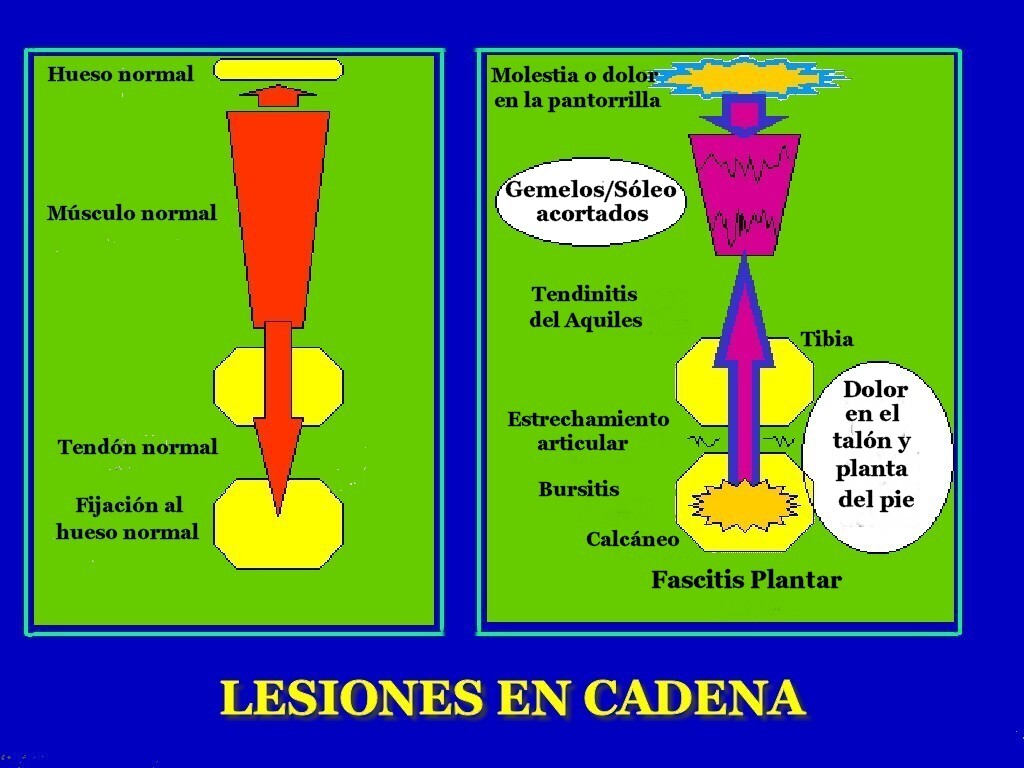
Knee/Hip/Back Pain - When the arch collapses in the foot, it triggers a series of compensations up the joint chain, leading to increased stress on the knee, pelvis and low back. Plantar fasciitis - This condition is characterized by heel pain, especially with the first few steps you take. The plantar fascia stretches as the arch falls, putting stress on the heel. Bunions - If you see a bony bump developing at the base of your big toe, you are likely developing a bunion. It may be swollen, red or painful when it rubs against your shoe. A flattened arch spreads the forefoot and causes the big toe to deviate toward the second toe. Shin splints - This term generally refers to pain anywhere along the shinbone. It is typically due to overuse and is aggravated after exercise and activity.

Diagnosis

Your doctor will ask about your symptoms and medical history. A physical and foot exam will be done. Flat feet can be diagnosed by appearance. To determine if the foot is rigid, you may be asked to do some simple tasks.

What is PES Planovalgus deformity?

Non Surgical Treatment

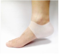
If you have fallen arches, but you are not experiencing any symptoms, then you probably do not need to seek treatment. If you are experiencing discomfort due to fallen arches, there are several treatment options. These treatment options include elevating the feet and applying ice to ease discomfort and reduce swelling, rest, exercises to stretch the feet, physical therapy, medication, such as anti-inflammatories, steroid injections and orthotic devices or customised arch supportsto wear in the shoes. If you have fallen arches and periodically experience pain related to that condition, it is a good idea to get orthotic devicesor custom arch supports, to wear in your shoes. The other treatment options, like medication and ice, will help to ease pain from fallen arches after you have already begun to experience pain. However, orthotic devices or(custom arch supports)can help to prevent pain from occurring at all. This preventative measure helps many people with fallen arches to avoid pain and prevent worsening of their condition. In severe cases of fallen arches, surgery may be required to correct the problem. You can also help to prevent pain and exacerbation of fallen arches by reducing your risk factors. If you are overweight, try to lose weight. Even a small weight loss can reduce the pressure on your feet significantly. If you are diabetic, manage your blood sugar as best as possible. Losing weight often also improves the condition of diabetics. You should also avoid high-impact activities, like running on the road, tennis, and sports that involve jumping. Try a gentler form of exercise, like swimming, instead. If you have fallen arches, orthotic devices or(custom arch supports)are an important component of your treatment and can help to prevent pain.
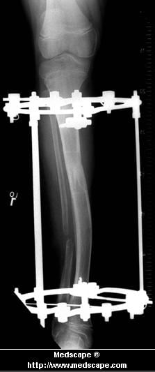
Surgical Treatment

Flat Foot

This is rare and usually only offered if patients have significant abnormalities in their bones or muscles. Treatments include joint fusion, reshaping the bones in the foot, and occasionally moving around tendons in the foot to help balance out the stresses (called tendon transfer).

After Care

Time off work depends on the type of work as well as the surgical procedures performed. . A patient will be required to be non-weight bearing in a cast or splint and use crutches for four to twelve weeks. Usually a patient can return to work in one to two weeks if they are able to work while seated. If a person's job requires standing and walking, return to work may take several weeks. Complete recovery may take six months to a full year. Complications can occur as with all surgeries, but are minimized by strictly following your surgeon's post-operative instructions. The main complications include infection, bone that is slow to heal or does not heal, progression or reoccurrence of deformity, a stiff foot, and the need for further surgery. Many of the above complications can be avoided by only putting weight on the operative foot when allowed by your surgeon.

Functional Leg Length Discrepancy Scoliosis

7/2/2017

 
Overview

Having one leg shorter than the other is a common physical condition. It has two primary causes--structural or functional problems. Structural differences in length can be the result of growth defect, previous injuries or surgeries. Functional differences in length can result from altered mechanics of the feet, knee, hip and/or pelvis. These altered mechanics from functional leg length discrepancy often stem from having an unbalanced foundation.Leg Length Discrepancy

Causes

Leg discrepancy can develop from a medical issue in any portion of the femur or tibia. One leg may lengthen, but leg shortening is much more common. Factors that can cause leg length discrepancy include inherited growth deficiencies. Infections. A bone infection can cause delayed growth in the affected limb. Injury. If your child breaks a leg, it may be shorter once it heals. This is most likely to happen if the fracture or break was complicated, an open fracture, or an injury that affected the growth plate near the end of the bone. Alternatively, a break can cause bones to grow faster after healing, making a leg longer. Tumors. Legg-Calve-Perthes disease. This is a condition that affects the ball (femoral head) of the hip joint. The femoral head may be friable and damage easily, sometimes leading to shortening of the thigh bone. Hemihypertrophy. In children with this condition, one side of the body grows more quickly than the other. Vascular malformations. These are abnormal clusters of veins and arteries that can form close to the bone and stimulate growth. Juvenile arthritis. Inflammation from arthritis can stimulate growth in the affected leg and cause discrepancy.

Symptoms

Often there are few or no symptoms prior to the age of 25-35. The most common symptom is chronic lower back pain, but also is frequently middle and upper back pain. Same-sided and repeated injury or pain to the hip, knee and/or ankle is also a hallmark of a long-standing untreated LLD. It is not uncommon to have buttock or radiating hip pain that is non-dermatomal (not from a disc) and tends to go away when lying down.

Diagnosis

On standing examination one iliac crest may be higher/lower than the other. However a physiotherapist, osteopath or chiropractor will examine the LLD in prone or supine position and measure it, confirming the diagnosis of structural (or functional) LLD. The LLD should be measured using bony fixed points. X-Ray should be taken in a standing position. The osteopath, physiotherapist or chiropractor will look at femoral head & acetabulum, knee joints, ankle joints.

Non Surgical Treatment

Non-surgical treatment can be effective. A shoe lift may be recommended if the leg length difference is less than 1 inch. More significant leg length discrepancies may require a surgical procedure. In children, surgical procedures are available to help make leg lengths more equal.

LLD Shoe Inserts

shoe lifts for men's shoes

Surgical Treatment

Surgeries for LLD are designed to do one of three general things ? shorten the long leg, stop or slow the growth of the longer or more rapidly growing leg, or lengthen the short leg. Stopping the growth of the longer leg is the most commonly utilized of the three approaches and involves an operation known as an epiphysiodesis , in which the growth plate of either the lower femur or upper tibia is visualized in the operating room using fluoroscopy (a type of real-time radiographic imaging) and ablated , which involves drilling into the region several times, such that the tissue is no longer capable of bone growth. Because the epiphyseal growth capabilities cannot be restored following the surgery, proper timing is crucial. Usually the operation is planned for the last 2 to 3 years of growth and has excellent results, with children leaving the hospital within a few days with good mobility. However, it is only appropriate for LLD of under 5cm.

Heel Painfulness All The Things You Might Want To Understand Heel Pain And Discomfort

6/29/2017

 
Overview

Pain On The Heel

Plantar fasciitis is the most common cause of heel pain, accounting for around four out of five cases. Plantar fasciitis is when the thick band of tissue that connects the heel bone with the rest of the foot (the plantar fascia) becomes damaged and thickened. Damage to the plantar fascia is thought to occur following, sudden damage - for example, damaging your heel while jogging, running or dancing; this type of damage usually affects younger people who are physically active, gradual wear and tear of the tissues that make up the plantar fascia - this usually affects adults who are 40 years of age or over.

Causes

Plantar fasciitis: It is the most common cause of heel pain. In this condition, the pain is more severe in the morning but becomes less painful as the day continues. It occurs due to tiny tears in the plantar fascia.The plantar faschia is a tissue band that connects the bottom of the heel bones to the ball of the foot and is involved in walking and running, giving spring to the step. If left untreated, the symptoms usually worsen and can lead to problems with the knee and hip and can cause back pain due to difficulty walking. Those who frequently stand or walk throughout the day or those who run are most likely to develop plantar fasciitis.

Symptoms

Depending on the specific form of heel pain, symptoms may vary. Pain stemming from plantar fasciitis or heel spurs is particularly acute following periods of rest, whether it is after getting out of bed in the morning, or getting up after a long period of sitting. In many cases, pain subsides during activity as injured tissue adjusts to damage, but can return again with prolonged activity or when excessive pressure is applied to the affected area. Extended periods of activity and/or strain of the foot can increase pain and inflammation in the foot. In addition to pain, heel conditions can also generate swelling, bruising, and redness. The foot may also be hot to the touch, experience tingling, or numbness depending on the condition.

Diagnosis

Your GP or podiatrist (a healthcare professional who specialises in foot care) may be able to diagnose the cause of your heel pain by asking about your symptoms and examining your heel and foot. You will usually only need further tests if you have additional symptoms that suggest the cause of your heel pain is not inflammation, such as numbness or a tingling sensation in your foot - this could be a sign of nerve damage in your feet and legs (peripheral neuropathy), your foot feels hot and you have a high temperature (fever) of 38C (100.4F) or above - these could be signs of a bone infection, you have stiffness and swelling in your heel - this could be a sign of arthritis. Possible further tests may include, blood tests, X-rays - where small doses of radiation are used to detect problems with your bones and tissues, a magnetic resonance imaging (MRI) scan or ultrasound scan, which are more detailed scans.

Non Surgical Treatment

Orthotics, by treating the cause of the problem, lead to the cure bette than 90% of the time. A small number of patients have waited so long that the plantar fascia has become thickened and filled with scar tissue and are not helped by ?conventional? means. Those are the patients that have, traditionally, required surgical treatment in which the plantar fascia is cut off the heel bone. Luckily, most surgery has been replaced by a relatively new mode of treatment, ESWT or Extracorporeal Shockwave Therapy. ESWT involves the application of multiple shockwaves to the diseased tendon or ligament and has an approximately 85 to 90% success rate. Keep in mind we are talking about this success rate in patients who are ?tough cases,? that is, already had the conventional treatment. The ESWT machines look like miniature renal lithtripsors (kidney stone crushers). There are virtually no side effects to ESWT other than the price as only about 30% of insurance companies are paying for it. They realize that it is less costly and safer than surgery but also know that many more people who would avoid surgery would have no problem getting ESWT so the volume of services would go up. You don?t have to live with painful heels.

Surgical Treatment

With the advancements in technology and treatments, if you do need to have surgery for the heel, it is very minimal incision that?s done. And the nice thing is your recovery period is short and you should be able to bear weight right after the surgery. This means you can get back to your weekly routine in just a few weeks. Recovery is a lot different than it used to be and a lot of it is because of doing a minimal incision and decreasing trauma to soft tissues, as well as even the bone. So if you need surgery, then your recovery period is pretty quick.

heel pain treatment

Prevention

Foot Pain

Before you get out of bed in the morning, and then periodically throughout the day, do the following exercises to increase flexibility and ease pain. Slowly flex your foot and toes to stretch the tissue on the bottom of your sore foot. Hold the stretch for 10 counts. Relax and repeat. Do gentle ankle rolls to keep the tissues around the ankle and on the back of the heel flexible. Sit on the edge of your bed and roll your foot back and forth over a tennis ball.

Leg Length Discrepancy Gait

6/26/2017

 
Overview

Surgery is another option. In some cases the longer extremity can be shortened, but a major shortening may weaken the muscles of the extremity. In growing children, lower extremities can also be equalized by a surgical procedure that stops the growth at one or two sites of the longer extremity, while leaving the remaining growth undisturbed. Your physician can tell you how much equalization can be attained by surgically halting one or more growth centers. The procedure is performed under X-ray control through very small incisions in the knee area. This procedure will not cause an immediate correction in length. Instead, the LLD will gradually decrease as the opposite extremity continues to grow and "catch up." Timing of the procedure is critical; the goal is to attain equal length of the extremities at skeletal maturity, usually in the mid- to late teens. Disadvantages of this option include the possibility of slight over-correction or under-correction of the LLD and the patient?s adult height will be less than if the shorter extremity had been lengthened. Correction of significant LLDs by this method may make a patient?s body look slightly disproportionate because of the shorter legs.Leg Length Discrepancy

Causes

Some limb-length differences are caused by actual anatomic differences from one side to the other (referred to as structural causes). The femur is longer (or shorter) or the cartilage between the femur and tibia is thicker (or thinner) on one side. There could be actual deformities in one femur or hip joint contributing to leg length differences from side to side. Even a small structural difference can amount to significant changes in the anatomy of the limb. A past history of leg fracture, developmental hip dysplasia, slipped capital femoral epiphysis (SCFE), short neck of the femur, or coxa vara can also lead to placement of the femoral head in the hip socket that is offset. The end-result can be a limb-length difference and early degenerative arthritis of the hip.

Symptoms

The most common symptom of all forms of LLD is chronic backache. In structural LLD the sufferer may also experience arthritis within the knee and hip are, flank pain, plantar fasciitis and metatarsalgia all on the side that is longer. Functional LLD sufferers will see similar conditions on the shorter side.

Diagnosis

The evaluation of leg length discrepancy typically involves sequential x-rays to measure the exact discrepancy, while following its progression. In addition, an x-ray of the wrist allows us to more carefully age your child. Skeletal age and chronological age do not necessarily equal each other and frequently a child's bone age will be significantly different than his or her stated age. Your child's physician can establish a treatment plan once all the facts are known: the bone age, the exact amount of discrepancy, and the cause, if it can be identified.

Non Surgical Treatment

Internal heel lifts: Putting a simple heel lift inside the shoe or onto a foot orthotic has the advantage of being transferable to many pairs of shoes. It is also aesthetically more pleasing as the lift remains hidden from view. However, there is a limit as to how high the lift can be before affecting shoe fit. Dress shoes will usually only accommodate small lifts (1/8"1/4") before the heel starts to piston out of the shoe. Sneakers and workboots may allow higher lifts, e.g., up to 1/2", before heel slippage problems arise. External heel lifts: If a lift of greater than 1/2" is required, you should consider adding to the outsole of the shoe. In this way, the shoe fit remains good. Although some patients may worry about the cosmetics of the shoe, it does ensure better overall function. Nowadays with the development of synthetic foams and crepes, such lifts do not have to be as heavy as the cork buildups of the past. External buildups are not transferable and they will wear down over time, so the patient will need to be vigilant in having them repaired. On ladies' high-heel shoes, it may be possible to lower one heel and thereby correct the imbalance.

LLD Insoles

what is a functional leg length discrepancy?

Surgical Treatment

Differences of an inch-and-a-half to two inches may require epiphysiodesis (adjusting the growth of the longer side) or acute shortening of the other side. Differences greater than 2.5 inches usually require a lengthening procedure. The short bone is cut and an external device is applied. Gradual lengthening is done over months to allow the muscles and nerves accommodate the new length.

What Is Mortons Neuroma

5/30/2017

 
Overview

intermetatarsal neuroma
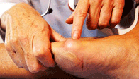
Patients with Morton?s neuroma present with pain in the forefoot, particularly in the ?ball? of the foot. However, not all pain in the forefoot is a Morton?s neuroma. In fact, most chronic pain in the forefoot is NOT the result of a Morton?s neuroma, but rather is from metatarsalgia - inflammation (synovitis) of the ?toe/foot? joints. The symptoms from Morton?s neuroma are due to irritation to the small digital nerves, as they pass across the sole of the foot and into the toes. Therefore, with a true Morton?s neuroma, it is not uncommon to have nerve-type symptoms, which can include numbness or a burning sensation extending into the toes. There are several interdigital nerves in the forefoot. The most common nerve to develop into a neuroma is between the 3rd and 4th toes. With a true neuroma, the pain should be isolated to just one or two toes.

Causes

Unfortunately, the cause of Morton?s Neuroma remains unknown to researchers. It is likely that a variety of factors may play a role in the development of this condition, including the presence of chronic pain conditions like fibromyalgia. Factors that may contribute to the development of Morton?s Neuroma include Wearing improperly fitting shoes can cause pressure on your foot, leading to swelling around the toe nerves. High heels are of particular concern as they cause a large amount of weight to be shifted to the ball of the foot. Repetitive activities like jogging, walking, and aerobics can also place a lot of pressure on the feet. This could lead to Morton?s Neuroma. Having a previous foot or muscle injury may cause you to hold your foot in a poor position when walking, contributing to nerve inflammation. Some people are just born with poorly shaped feet. People with extremely low arches or "flat feet" may suffer from Morton?s Neuroma more than others.

Symptoms

A Morton's neuroma usually causes burning pain, numbness or tingling at the base of the third, fourth or second toes. Pain also can spread from the ball of the foot out to the tips of the toes. In some cases, there also is the sensation of a lump, a fold of sock or a "hot pebble" between the toes. Typically, the pain of a Morton's neuroma is relieved temporarily by taking off your shoes, flexing your toes and rubbing your feet. Symptoms may be aggravated by standing for prolonged periods or by wearing high heels or shoes with a narrow toe box.

Diagnosis

A thorough subjective and objective examination from a physiotherapist is usually sufficient to diagnose a Morton's neuroma. Investigations such as an X-ray, ultrasound, MRI, CT scan or bone scan may sometimes be used to assist with diagnosis, assess the severity of the injury and rule out other conditions.

Non Surgical Treatment

Treatment for Morton's neuroma may depend on several factors, including the severity of symptoms and how long they have been present. The earlier on the condition is diagnosed, the less likely surgery is required. Doctors will usually recommend self-help measures first. These may include resting the foot, massaging the foot and affected toes. Using an ice pack on the affected area (skin should not be directly exposed to ice, the ice should be in a container or wrapped in something) Changing footwear, wearing wide-toed shoes, or flat (non high-heeled) shoes. Trying arch supports (orthotic devices). A type of padding that supports the arch of the foot, removing pressure from the nerve. The doctor may recommend a custom-made, individually designed shoe-insert, molded to fit the contours of the patient's foot. There are several OTC (over the counter, non-prescription) metatarsal pads or bars available which can be placed over the neuroma. Taking over-the-counter, non-prescription painkilling medications. Modifying activities, avoiding activities which put repetitive pressure on the neuroma until the condition improves. Bodyweight management,if the patient is obese the doctor may advise him/her to lose weight. A significant number of obese patients with foot problems, such as flat feet, who successfully lose weight experience considerable improvement of symptoms.Morton neuroma

Surgical Treatment

When conservative measures are unsuccessful, surgery can be a good choice in the treatment of Morton's neuroma. The operation for Morton's neuroma does not require an overnight hospital stay. The anesthetic used is an ankle block, which completely numbs the foot during the surgery. The physician removes the neuroma from an incision made on the top of the foot between the involved metatarsal heads. The nerve to the interspace is exposed and cut next to the metatarsal heads.

Leg Length Discrepancy And Shoe Lifts

2/23/2016

 
There are two unique variations of leg length discrepancies, congenital and acquired. Congenital means that you are born with it. One leg is anatomically shorter in comparison to the other. As a result of developmental periods of aging, the brain picks up on the step pattern and identifies some difference. The human body typically adapts by tilting one shoulder to the "short" side. A difference of less than a quarter inch is not grossly abnormal, does not need Shoe Lifts to compensate and normally doesn't have a profound effect over a lifetime.

Shoe Lift

Leg length inequality goes largely undiscovered on a daily basis, yet this issue is simply remedied, and can reduce a number of cases of back ache.

Treatment for leg length inequality typically consists of Shoe Lifts . These are generally low-priced, typically priced at less than twenty dollars, compared to a custom orthotic of $200 or more. When the amount of leg length inequality begins to exceed half an inch, a whole sole lift is generally the better choice than a heel lift. This prevents the foot from being unnecessarily stressed in an abnormal position.

Chronic back pain is the most widespread health problem afflicting people today. Around 80 million people are affected by back pain at some stage in their life. It's a problem which costs employers millions annually as a result of time lost and productivity. Innovative and more effective treatment methods are always sought after in the hope of minimizing the economical influence this issue causes.

Leg Length Discrepancy Shoe Lifts

Men and women from all corners of the world suffer from foot ache due to leg length discrepancy. In most of these situations Shoe Lifts might be of very useful. The lifts are capable of decreasing any discomfort in the feet. Shoe Lifts are recommended by numerous specialist orthopaedic physicians.

So as to support the body in a well-balanced manner, feet have a significant function to play. Despite that, it can be the most neglected zone in the body. Some people have flat-feet meaning there may be unequal force placed on the feet. This causes other body parts like knees, ankles and backs to be impacted too. Shoe Lifts ensure that suitable posture and balance are restored.

Leg Length Discrepancy And Shoe Lifts

2/22/2016

 
There are actually two different types of leg length discrepancies, congenital and acquired. Congenital implies you are born with it. One leg is structurally shorter in comparison to the other. Through developmental periods of aging, the human brain picks up on the stride pattern and recognizes some variance. Your body typically adapts by tilting one shoulder to the "short" side. A difference of under a quarter inch isn't really abnormal, require Shoe Lifts to compensate and in most cases doesn't have a serious effect over a lifetime.

Leg Length Discrepancy Shoe Lifts

Leg length inequality goes largely undiscovered on a daily basis, however this condition is simply fixed, and can reduce a number of cases of lower back pain.

Therapy for leg length inequality typically consists of Shoe Lifts. These are very inexpensive, typically priced at under twenty dollars, compared to a custom orthotic of $200 or higher. When the amount of leg length inequality begins to exceed half an inch, a whole sole lift is generally the better choice than a heel lift. This prevents the foot from being unnecessarily stressed in an abnormal position.

Chronic back pain is the most common condition afflicting people today. Around 80 million men and women have problems with back pain at some stage in their life. It is a problem which costs employers millions yearly on account of time lost and production. Innovative and more effective treatment methods are always sought after in the hope of decreasing the economic impact this condition causes.

 <a href="http://tallervictoria5677.pen.io">Shoe Lifts</a>

Men and women from all corners of the world suffer from foot ache as a result of leg length discrepancy. In these cases Shoe Lifts can be of very helpful. The lifts are capable of alleviating any discomfort in the feet. Shoe Lifts are recommended by many specialist orthopaedic physicians.

In order to support the body in a healthy and balanced manner, the feet have a critical function to play. In spite of that, it can be the most neglected area in the body. Many people have flat-feet which means there may be unequal force placed on the feet. This will cause other body parts such as knees, ankles and backs to be impacted too. Shoe Lifts guarantee that the right posture and balance are restored.

Do Bunions Demand Surgery

6/18/2015

 
Overview
Bunion Pain A bunion is the enlargement of the joint at the base of the big toe. It occurs as a result of the misalignment of the bones of the big toe. This leads to stretching of the ligaments and tendons around the big toe joint and causes soft tissue over the joint to become inflamed and painful. There may be additional bone formation (exostosis) in the joint and the skin around the joint may become red and tender. Over time the cartilage in the joint can break down, leading to arthritis.

Causes
Contributing factors may include excessive foot pronation, wearing tight and pointed-toe shoes, and occasionally trauma. Joint misalignment causes osteoarthritis with cartilage erosion and exostosis formation, resulting in joint motion being limited (hallux limitus) or eliminated (hallux rigidus). In late stages, synovitis occurs, causing joint swelling. In reaction to pressure from tight shoes, an adventitious bursa can develop medial to the joint prominence, which can become painful, swollen, and inflamed.

Symptoms
Your bunion may not cause any symptoms. Or you may have pain in your big toe, red or irritated skin over the bunion, and swelling at the base of the big toe. The big toe may point toward the other toes and cause problems in other toes, such as hammer toe . A bunionette can cause similar symptoms at the base of the little toe.

Diagnosis
When an x-ray of a bunion is taken, there is usually angulation between the first metatarsal bone and the bones of the big toe. There may also be angulation between the first and second metatarsal bones. These angular irregularities are the essence of most bunions. In general, surgery for bunions aims to correct such angular deformities.

Non Surgical Treatment
The non-invasive treatments for bunions are many and include changes in footwear, icing the sore area, over the counter pain medications, orthotic shoe inserts, and weight management. If these conservative measures fail to arrest your pain and discomfort, your foot and ankle surgeon may recommend a bunionectomy or similar surgical procedure, depending on your condition. Bunions Hard Skin

Surgical Treatment
When a surgeon cuts and repositions a bone, it is referred to as an osteotomy. There are two basic techniques used to perform an osteotomy to realign the first metatarsal. In some cases, the far end of the bone is cut and moved laterally (called a distal osteotomy). This effectively reduces the angle between the first and second metatarsal bones. This type of procedure usually requires one or two small incisions in the foot. Once the surgeon is satisfied with the position of the bones, the osteotomy is held in the desired position with one, or several, metal pins. Once the bone heals, the pin is removed. The metal pins are usually removed between three and six weeks following surgery. In other situations, the first metatarsal is cut at the near end of the bone (called a proximal osteotomy). This type of procedure usually requires two or three small incisions in the foot. Once the skin is opened the surgeon performs the osteotomy. The bone is then realigned and held in place with metal pins until it heals. Again, this reduces the angle between the first and second metatarsal bones. Realignment of the big toe is then done by releasing the tight structures on the lateral, or outer, side of the first MTP joint. This includes the tight joint capsule and the tendon of the adductor hallucis muscle. This muscle tends to pull the big toe inward. By releasing the tendon, the toe is no longer pulled out of alignment. The toe is realigned and the joint capsule on the side of the big toe closest to the other toe is tightened to keep the toe straight, or balanced. Once the surgeon is satisfied that the toe is straight and well balanced, the skin incisions are closed with small stitches. A bulky bandage is applied to the foot before you are returned to the recovery room.

Prevention
Shoes that possess tapering toe boxes should be avoided if you have a bunion, as narrow toe boxes will hasten the progression of your bunion deformity. In some cases, conservative measures, including switching to appropriate footwear, may not have the desired effect, and your podiatrist may recommend for you a surgical procedure known as a bunionectomy.

What Is Calcaneal Apophysitis?

5/23/2015

 
Overview

Sever's disease is the most common cause of heel pain in the growing child. It is due to overuse and repetitive stress of the Achilles tendon where it attaches to the heel bone.

Causes

When a baby is born, most of the bones are still cartilage with only some starting to develop into bone. When the heel (calcaneus) starts to develop bone, there is generally one large area of development that starts in the center of the cartilage heel. This area of bone spreads to 'fill up' the cartilage. Another area of bone development (ossification) occurs at the back of the heel bone. These two areas of developing bone will have an area of cartilage between them, this is how the bone grows in size. At around age 16, when growth is nearly complete, these two bony areas fuse together. Sever's disease or calcaneal apophysitis is usually considered to be due to damage or a disturbance in this area of growth.

Symptoms

Unilateral or bilateral heel pain. Heel pain during physical exercise, especially activities that require running or jumping. Increased pain level after exercise. A tender swelling or bulge on the heel that is painful on touch. Limping. Calf muscle stiffness first thing in the morning.

Diagnosis

A physical exam of the heel will show tenderness over the back of the heel but not in the Achilles tendon or plantar fascia. There may be tightness in the calf muscle, which contributes to tension on the heel. The tendons in the heel get stretched more in patients with flat feet. There is greater impact force on the heels of athletes with a high-arched, rigid foot.

Non Surgical Treatment

Treatment depends on the severity of the condition, but may include relative rest and modified activity, a physiotherapist can help work out what, and how much, activity to undertake. Cold packs, apply ice or cold packs to the back of the heels for around 15 minutes after any physical activity, including walking. Shoe inserts, small heel inserts worn inside the shoes can take some of the traction pressure off the Achilles tendons. This will only be required in the short term. Medication, pain-relieving medication may help in extreme cases, but should always be combined with other treatment and following consultation with your doctor). Anti-inflammatory creams are also an effective management tool. Splinting or casting, in severe cases, it may be necessary to immobilise the lower leg using a splint or cast, but this is rare. Time, generally the pain will ease in one to two weeks, although there may be flare-ups from time to time. Correction of any biomechanical issues, a physiotherapist can identify and discuss any biomechanical issues that may cause or worsen the condition. Education on how to self-manage the symptoms and flare-ups of Sever?s disease is an essential part of the treatment.

Exercise

For children with Sever's disease, it is important to habitually perform exercises to stretch the hamstrings, calf muscles, and the tendons on the back of the leg. Stretching should be performed 2-3 times a day. Each stretch should be performed for 20 seconds, and both legs should be stretched, even if the pain is only in one heel. Heel cups or an inner shoe heel lifts are often recommended for patient suffering from Sever's disease. Wearing running shoes with built in heel cups can also decrease the symptoms because they can help soften the impact on the heel when walking, running, or standing.

Acquired Flat Foot Tibialis Posterior Pain

4/27/2015

 
Overview
A painful flat foot, or adult acquired flatfoot deformity, is a progressive collapsing of the arch of the foot that occurs as the posterior tibial tendon becomes insufficient due to various factors. Early stages may present with only pain along the posterior tibial tendon whereas advanced deformity usually results in arthritis and rigidity of the rearfoot and ankle. Flat Foot

Causes
As discussed above, many health conditions can create a painful flatfoot. Damage to the posterior tibial tendon is the most common cause of AAFD. The posterior tibial tendon is one of the most important tendons of the leg. It starts at a muscle in the calf, travels down the inside of the lower leg and attaches to the bones on the inside of the foot. The main function of this tendon is to hold up the arch and support your foot when you walk. If the tendon becomes inflamed or torn, the arch will slowly collapse. Women and people over 40 are more likely to develop problems with the posterior tibial tendon. Other risk factors include obesity, diabetes, and hypertension. Having flat feet since childhood increases the risk of developing a tear in the posterior tibial tendon. In addition, people who are involved in high impact sports, such as basketball, tennis, or soccer, may have tears of the tendon from repetitive use. Inflammatory arthritis, such as rheumatoid arthritis, can cause a painful flatfoot. This type of arthritis attacks not only the cartilage in the joints, but also the ligaments that support the foot. Inflammatory arthritis not only causes pain, but also causes the foot to change shape and become flat. The arthritis can affect the back of the foot or the middle of foot, both of which can result in a fallen arch.

Symptoms
The symptoms of PTTD may include pain, swelling, a flattening of the arch, and inward rolling of the ankle. As the condition progresses, the symptoms will change. For example, later, as the arch begins to flatten, there may still be pain on the inside of the foot and ankle. But at this point, the foot and toes begin to turn outward and the ankle rolls inward. As PTTD becomes more advanced, the arch flattens even more and the pain often shifts to the outside of the foot, below the ankle. The tendon has deteriorated considerably and arthritis often develops in the foot. In more severe cases, arthritis may also develop in the ankle. Symptoms, which may occur in some persons with flexible flatfoot, include. Pain in the heel, arch, ankle, or along the outside of the foot. ?Turned-in? ankle. Pain associated with a shin splint. General weakness / fatigue in the foot or leg.

Diagnosis
Diagnostic testing is often used to diagnose the condition and help determine the stage of the disease. The most common test done in the office setting are weightbearing X-rays of the foot and ankle. These assess joint alignment and osteoarthritis. If tendon tearing or rupture is suspected, the gold standard test would be MRI. The MRI is used to check the tendon, surrounding ligament structures and the midfoot and hindfoot joints. An MRI is essential if surgery is being considered.

Non surgical Treatment
Icing and anti-inflammatory medications can reduce inflammation and physical therapy can strengthen the tibial tendon. Orthotic inserts that go inside your shoes are a common way to treat and prevent flatfoot pain. Orthotics control the position of the foot and alleviate areas of pressure. In some cases immobilization in a cast or walking boot is necessary to relieve symptoms, and in severe cases surgery may be required to repair tendon damage. Flat Foot

Surgical Treatment
In cases of PTTD that have progressed substantially or have failed to improve with non-surgical treatment, surgery may be required. For some advanced cases, surgery may be the only option. Surgical treatment may include repairing the tendon, tendon transfers, realigning the bones of the foot, joint fusions, or both. Dr. Piccarelli will determine the best approach for your specific case. A variety of surgical techniques is available to correct flexible flatfoot. Your case may require one procedure or a combination of procedures. All of these surgical techniques are aimed at relieving the symptoms and improving foot function. Among these procedures are tendon transfers or tendon lengthening procedures, realignment of one or more bones, or insertion of implant devices. Whether you have flexible flatfoot or PTTD, to select the procedure or combination of procedures for your particular case, Dr. Piccarelli will take into consideration the extent of your deformity based on the x-ray findings, your age, your activity level, and other factors. The length of the recovery period will vary, depending on the procedure or procedures performed.
Powered by Create your own unique website with customizable templates.